The Enamel Review · Technology
How CT Resolution Shapes Bone-Implant Predictions
Scan resolution and stiffness rules can quietly rewrite what a computer predicts at the bone-implant interface.
In a natural, human-quality voice · about 4 min · free when you register
Read aloud · 0:00 / 4:00 · the text follows along
Computers have become quiet partners in implant dentistry. A scan can be turned into a mesh, a mesh into a stress map, and a map into a conversation about risk. The promise is precision. Every prediction is only as faithful as the pictures and the material rules that feed it. Researchers writing in Computer Methods and Programs in Biomedicine examine two of those inputs—CT image resolution and the way bone stiffness is assigned—and consider how they shape biomechanical forecasts at the bone-implant interface, where load crosses into living bone.
Why the bone-implant interface is a modeling problem
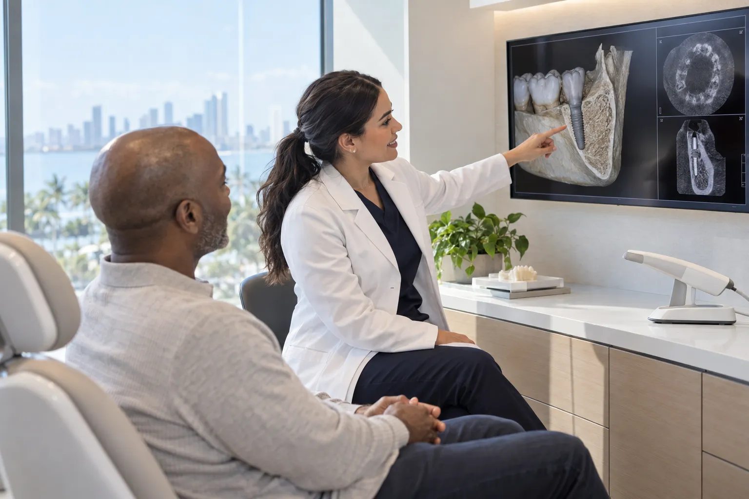
When an implant is loaded, force does not vanish into the fixture. It crosses into bone. That crossing is not a single number. Cortical plates, trabecular struts, the tightness of contact, and the direction of bite all matter. Finite-element models try to stand in for that complexity so a clinician experienced in full-arch planning can see where strain might concentrate. They are hypotheses rendered in color. If the mesh is too coarse, a peak can be smoothed away. If bone is treated as a uniform block, a weak region can be assigned the stiffness of a dense one. Those choices are easy to hide inside software. They are harder to defend once a patient is in the chair.
What CT resolution actually captures
Computed tomography does not photograph bone. It samples it. Voxel size sets the smallest feature the scan can represent without blur. Trabeculae in the jaws are thin. When voxels are large relative to those struts, the image averages marrow and mineral into a gray that looks like a solid of middling density. The computer then builds a geometry from that average. Fine threads of bone may disappear. At the implant surface, where contact is local, those errors are not academic. They change how load is thought to spread. Higher resolution is not automatically better for every clinical workflow. It can mean larger files and a different radiation burden depending on protocol. The scientific question is whether the extra detail moves the prediction at the interface enough to matter for planning, or whether it mainly changes the look of a figure. That is the kind of sensitivity work computational papers exist to do. Readers should treat resolution as a setting with consequences, not as a badge of quality.
How material modeling rewrites the same scan
Even a crisp CT volume still needs a rule that turns gray values into mechanical properties. One common path converts Hounsfield units toward density and then toward elastic modulus. Another assigns a few discrete materials—cortical, cancellous, implant alloy—and stops there. Heterogeneous maps can look more realistic. Homogeneous maps are simpler to check. Neither is the bone. Both are models of bone. At the interface, the difference shows up as stress and strain that may cluster or spread, and as micromotion estimates that may rise or fall. Anisotropy, the fact that bone is stiffer along some directions than others, is often left out. Damage, remodeling, and the biology of osseointegration are left out almost always. A related Enamel Review piece on a biomimetic bilayer membrane for periodontal regeneration reminds readers that the living interface is a tissue problem, not only a mesh problem. Computational studies that vary material laws are useful precisely because they show how much the story depends on those laws. They do not crown a single law as truth.
What a prediction is—and is not
Patients sometimes meet these images as reassurance: colored plots, arrows, a sense that the plan has been proven. Dentists meet them as decision support. The honest word for what a well-built model offers is association between assumed conditions and calculated fields, not a guarantee of clinical outcome. The FDA reviews many imaging devices and implant systems on safety and performance evidence of a different kind; a journal paper about mesh sensitivity is not a substitute for that process, and it is not a treatment recommendation. NIDCR public education on implants frames them as options that depend on bone, general health, and maintenance—not on a particular software pipeline. Cochrane-style systematic reviews, when they exist for implant questions, look at trials in people. A methods paper looks at numbers in a computer. Those two kinds of evidence answer different questions. For full-mouth rehabilitation, including same-day full arch workflows that rely on in-house milling and quick turnover where a clinic qualifies, the scan-to-model chain is part of the craft. It should be discussed as craft: what was scanned, at what resolution, how bone was assigned, and which loading cases were run.
If you are a patient looking at a colorful implant simulation, ask what the colors assume. If you are a dentist reading a biomechanical paper, look first at voxel size, material assignment, and contact rules. The takeaway is modest and useful. Better pictures and more careful material models can change the numbers at the bone-implant boundary. They do not replace an exam, a conversation about health, or the slow biology of bone that no mesh fully owns.
Comments
Comments publish after a one-click email verification — every voice here is a real one. Comments are reviewed before they appear.
Loading comments…