The Enamel Review · Artificial Intelligence
Automated landmarks on 3D dental models: how it works
A new study in Computer Methods and Programs in Biomedicine flattens 3D dental scans into images, then lets a two-stage network find the landmarks that model analysis depends on.
In a natural, human-quality voice · about 4 min · free when you register
Read aloud · 0:00 / 4:00 · the text follows along
Model analysis is one of the oldest quiet rituals in dentistry. Someone takes a cast — plaster once, a mesh now — and measures it: intercanine and intermolar widths, arch length, tooth dimensions, the arch-perimeter arithmetic that turns into a crowding estimate. The measurements are unglamorous and consequential, because they sit underneath decisions about extraction, expansion, space management, and the sequencing of complex restorative work. They are also slow, hand-driven, and variable between operators. A paper published in Computer Methods and Programs in Biomedicine (2026) proposes a way to automate the landmark-finding step on 3D digital dental models using a projection-based two-stage cascade convolutional neural network — an approach worth understanding, because the design choice it makes is the interesting part.
What does a projection-based cascade actually do?
A digital dental model is a triangle mesh: hundreds of thousands of vertices with no fixed ordering and no guaranteed orientation. That irregularity is exactly what conventional convolutional networks dislike. Networks that operate directly on meshes and point clouds exist, but they are computationally expensive and hungry for annotated data, which is scarce in dentistry.
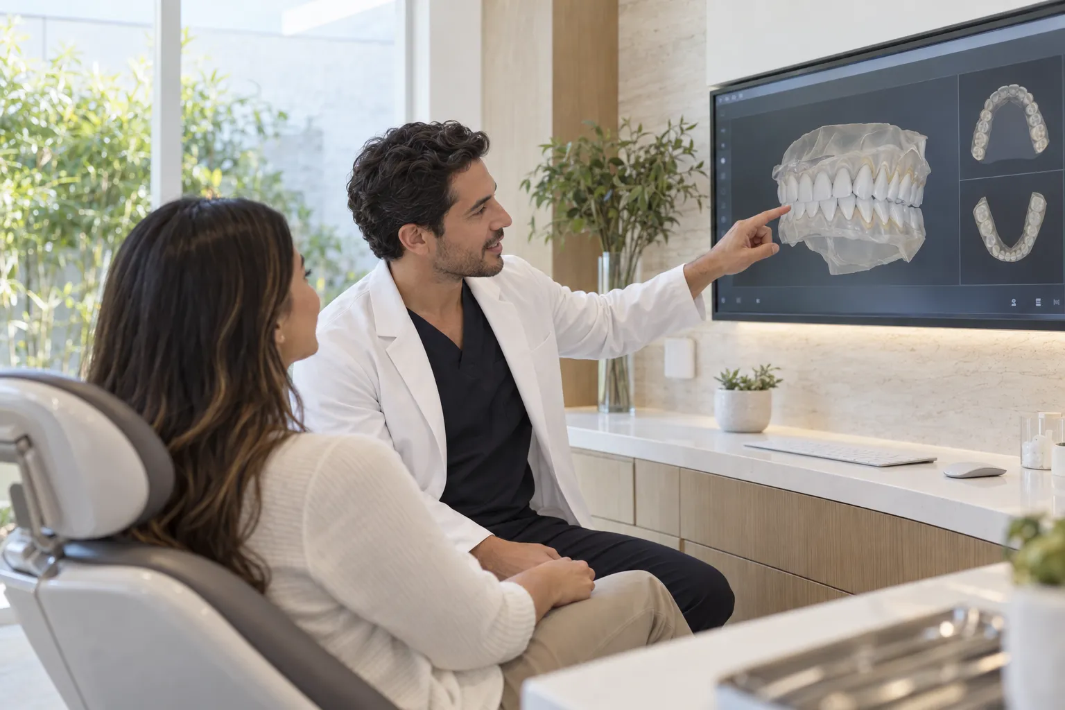
The projection strategy sidesteps the problem. Instead of learning on the mesh, the model is rendered into one or more 2D images — typically depth or curvature maps viewed from defined directions — so that the mature machinery of image-based convolutional networks can be applied. The cascade then works coarse-to-fine: a first stage locates approximate landmark regions across the whole arch, and a second stage crops in on those regions and refines each position at higher resolution. Refined 2D coordinates are mapped back onto the mesh surface, and the resulting 3D landmarks feed the standard model-analysis measurements.
The logic is the same as a clinician’s: orient, scan the whole arch, then look closely at the cusp tip or the contact point that matters. Two stages beat one because a single network asked to be both global and precise usually ends up being neither.
Why is model analysis a sensible first target for automation?
Automation earns its place where a task is repetitive, rule-governed, and already measured against an imperfect human standard. Model analysis qualifies on all three counts. The definitions are largely explicit — mesiobuccal cusp tips, canine cusps, incisal midpoints, gingival zeniths — and the arithmetic downstream is deterministic. Once landmarks are placed, arch widths and Bolton-style tooth-size comparisons are simple subtraction and division.
There is also a practical reason. Intraoral scanning has moved digital models from the plaster shelf into routine records, so the input data already exists in most offices that have adopted a scanner. Automated landmarking does not require a new acquisition step; it asks something more of files that are being captured anyway. That matters for diagnostic workflows and for planning that crosses disciplines — orthodontic space analysis, prosthetically driven implant planning, or the sequencing of a full-mouth rehabilitation, where arch form and tooth proportion inform the entire treatment sequence.
Where do automated landmarks go wrong?
Neutrally stated: the study reports its own accuracy figures against human annotation, and readers who care about the numbers should read the paper rather than a summary of it. The more durable point concerns the failure modes, which are predictable in any projection-based system.
Projection loses information. Surfaces hidden from the chosen viewing direction — deep lingual contours, steep palatal walls, structures shadowed by crowded or rotated teeth — are compressed or absent in the rendered image. Landmarks in those zones are the ones most likely to drift. Missing teeth, mixed dentition, orthodontic attachments, restorations with unusual anatomy, and scan artifacts at the gingival margin all shift the input away from whatever distribution the network was trained on.
And the reference standard is itself human. When an algorithm agrees closely with expert annotation, the finding is an association between two measurement processes, not proof that either is correct. Agreement studies of this kind describe reproducibility; they do not establish that an automated arch-length value changes a treatment decision for the better. That would require prospective work of a different design, and neither this paper nor any single paper should be read as settling it.
What should a reader take from this?
For patients, the honest framing is modest and reassuring: software of this type speeds up a measurement step that has always been part of careful record-keeping. It does not replace a diagnosis, and it does not decide anything. For dentists evaluating such tools, the questions are the familiar ones — what data was the network trained on, which landmarks degrade first, is there a visible confidence signal, and how easily can a clinician override a misplaced point without rebuilding the analysis from scratch.
The practical takeaway is simple. If your records include a digital scan, ask how the measurements derived from it were produced and who checked them. An automated landmark is a starting point that a trained eye should confirm; used that way, it saves time without quietly taking over judgment. Regulatory status for clinical software is determined by agencies such as the FDA, and research-grade methods described in the literature are not, by themselves, cleared clinical products.
Comments
Comments publish after a one-click email verification — every voice here is a real one. Comments are reviewed before they appear.
Loading comments…